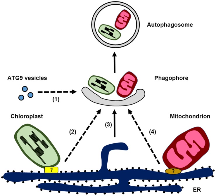
FIGURE 1.
Schematic illustration of autophagosome biogenesis in plant cells, highlighting the possible membrane sources for phagophore formation: (1) ATG9 vesicles, (2) endoplasmic reticulum (ER)-chloroplast contact site, (3) ER, and (4) ER-mitochondria contact site. Potential protein complex responsible for the ER-chloroplast contact site and ER-mitochondria contact site are labeled with the question mark.

