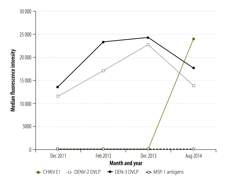
Fig. 1.
Fluorescence intensities recorded in multiplex bead assays based on antigens representing chikungunya and dengue viruses and Plasmodium falciparum, Haiti, 2011–2014
CHIKV E1: chikungunya virus envelope 1; DVLP: dengue virus-like particle; MSP-1: merozoite surface protein 1.
Notes: The assays were used to investigate blood spots from a longitudinal cohort of 61 children. The intensities shown are each the result of subtracting background fluorescence, from wells containing no primary antibody, from the mean of the median fluorescence intensities of duplicate wells. The three P. falciparum antigens tested – i.e. 19-kDa, 42-kDa and 42-kDa fractions of the MSP-1 clones 3D7, 3D7 and FVO, respectively, gave almost identical results.

