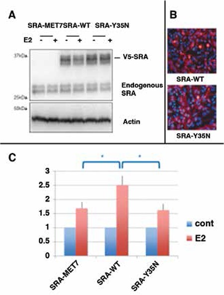
Figure 4. Functional analyses of SRA1 mutant p.Y35N A) Levels of SRAP expression protein extracts of transfected HeLa cells. Equal volumes of luciferase assay extracts from HeLa cells transfected with Estrogen Receptor-α (ESR1ESR1) and PS2-ERE luciferase reporter plasmids, and either control (SRA-MET7), wild-type (SRA-WT), or mutant (SRA-Y35N) SRA plasmids, were subjected to western blot analysis using anti-SRAP (743, Bethyl Laboratories) and anti-Actin antibodies (Abcam). Shown is a representative blot displaying equal levels of exogenous V5-epitope tagged SRAP (~35kDa) products in both SRA-WT and SRA-Y35N but not control SRA-MET7 transfected cell lysates. B) Pancellular localization of wild-type versus Y35N SRAP. HeLa cells were transiently transfected with either V5-epitope tagged SRA-WT or SRA-Y35N constructs and exogenous SRAP (Red) expression was observed by indirect immunofluorescent microscopy using anti-V5 (Life Technologies) primary antibody followed by anti-Mouse-Alexa555 (Life Technologies). Cells were counterstained with Dapi to visualize nuclei (Blue). C) SRA-Y35N mutation results in impaired estradiol induced ESR1 transactivation of PS2-ERE luciferase reporter. HeLa cells were co-transfected with Estrogen Receptor-α (ESR1), PS2-ERE luciferase reporter, and either control (SRA-MET7), wild-type (SRA-WT), or mutant (SRA-Y35N) SRA plasmids 24 h prior to being treated with estradiol (+E2) or ethanol (cont). Data were normalized as detailed in the Materials and Methods section. Error bars represent standard deviation for n=4. Unpaired 2 tailed student’s t-test was performed to test for significant difference among different conditions (*represents p<0.05).

