FIGURE 2.
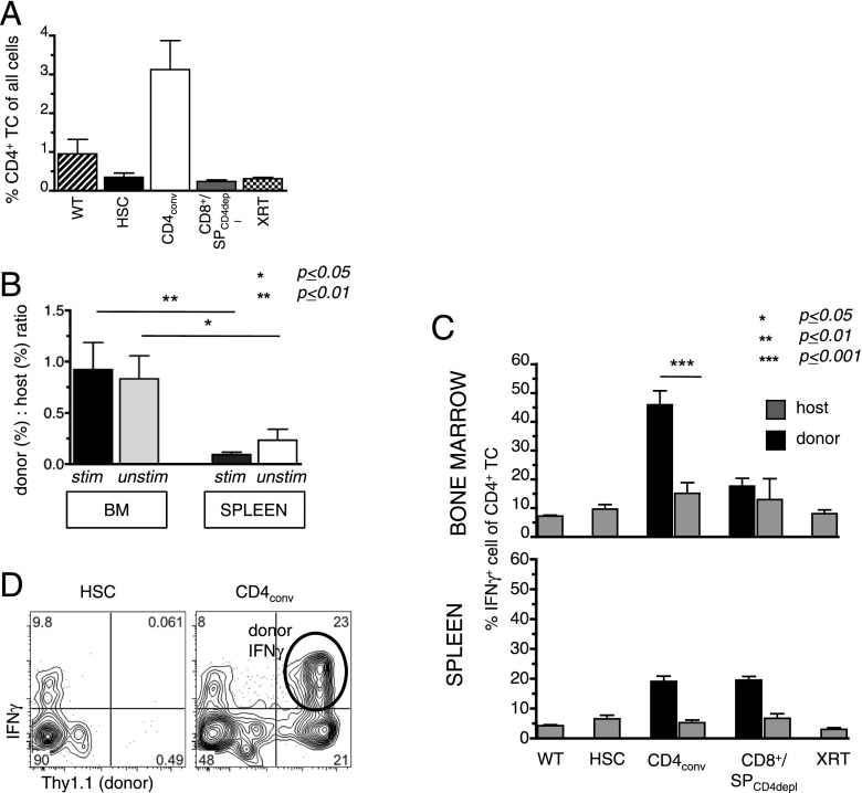
Donor CD4+ T cells induce Th1 immune reactions in the marrow post-HCT. As in Fig. 1A, BALB.K mice were transplanted with AKR/J (both H2k) KTLS-HSCs alone or in combination with CD4conv T cells (CD4+CD25−), or CD8+/SPCD4depl (both groups were compiled as one group). BM and spleens of recipients were harvested on day 14 for measurement of cell content and intracellular cytokines. (A) BM cells contained higher proportions of CD4+ T cells in HSC+CD4conv recipients (n = 7) compared with the other groups (WT: n = 2; HSC alone: n = 6; SPCD4depl: n = 4; XRT only: n = 4), as determined by FACS. (B) The ratio of donor to host contribution within the CD4+ T cells was calculated by % donor CD4+/% host CD4+ T cells of all live cells. In HSC+CD4conv recipients, donor-host ratios were significantly higher in the BM as compared with the spleen for both PMA-stimulated and unstimulated T cells (n = 11). (C) Compiled data of % IFN-γ+ cells of all BM and spleen CD4+ T cells (after 6-h PMA stimulation) in WT controls (n = 2), recipients of HSC only (n = 6), HSC+CD4conv (n = 7), HSC+CD8+/SPCD4depl (n = 4), or XRT controls (n = 4). Percentages of donor- and host-derived IFN-γ+ are shown for transplanted groups. (D) Representative FACS plots gated on BM CD4+ T cells in recipients of HSCs and HSC+CD4conv show intracellular IFN-γ+ expression of donor (Thy1.1+) and host (Thy1.1−) CD4+ T cells. *p ≤ 0.05, **p ≤ 0.01, ***p ≤ 0.001.

