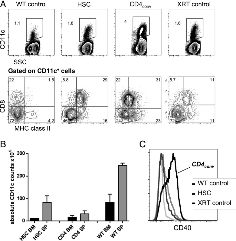
FIGURE 3.
Higher levels of activated CD11c+ cells are found in the marrow of CD4+ T cell recipients. (A) Representative FACS plots show the proportion of CD11c+ cells of all live cells in the BM of WT mice, recipients of HSCs, recipients of HSC+CD4conv, and XRT controls on day 7 post-HCT (upper panels) and their coexpression of CD8+ and MHC II (lower panels, gated on CD11c+). (B) Absolute counts of CD11c+ cells in the BM (black bars) and spleen (gray bars) of sublethally irradiated BALB.K recipients of AKR/J HSCs or HSC+CD4+ at 2 wk post-HCT as compared with WT controls, showing no significant differences in absolute cell counts in both experimental groups, but higher numbers in the WT mice. (C) Representative FACS histograms showing CD11c+ cells had increased CD40 expression in HSC+CD4conv recipients compared with controls at 1 wk post-HCT.

