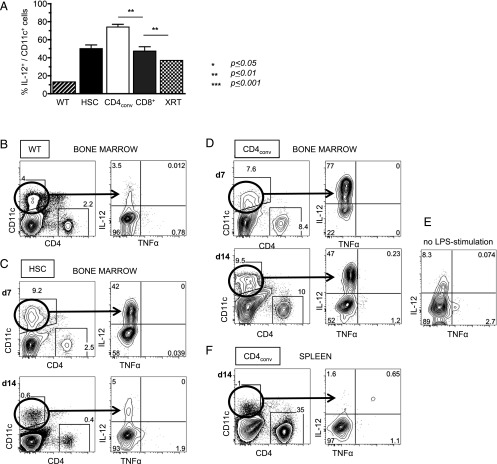
FIGURE 4.
IL-12–secreting CD11c+ DCs persist in recipients of HSC+CD4+ T cells. (A) Compiled data on IL-12 expression of CD11c+ DCs in LPS-stimulated BM at day (d) 7 post-HCT showing significantly higher levels of IL-12 in HSC+CD4conv recipients as compared with recipients of HSCs or HSC+CD8+ T cells. (B–F) Representative FACS plots of WT BM cells (B); BM from HSC recipients on d7 and d14 post-HCT (C); BM from HSC+CD4conv recipients on d7 and d14 post-HCT (D) including an unstimulated control from the same animal (E); spleen from HSC+CD4conv recipients on d14 post-HCT (F) after 14-h in vitro LPS stimulation, displaying CD11c+ DCs and their baseline IL-12 expression. (C) In HSC recipients, DCs expanded upon LPS stimulation and expressed IL-12 on d7 post-HCT but normalized by d14. (D) In recipients of HSC+CD4conv, DC expansion and IL-12 expression persisted through d14 post-HCT. (E) In recipients of HSC+CD4conv, IL-12 expression was also detectable without LPS stimulation. (F) In recipients of HSC+CD4conv, no increased IL-12 expression was detectable in the spleen. The number of experimental animals was n = 3–5 per time point per group. **p ≤ 0.01.

