FIGURE 5.
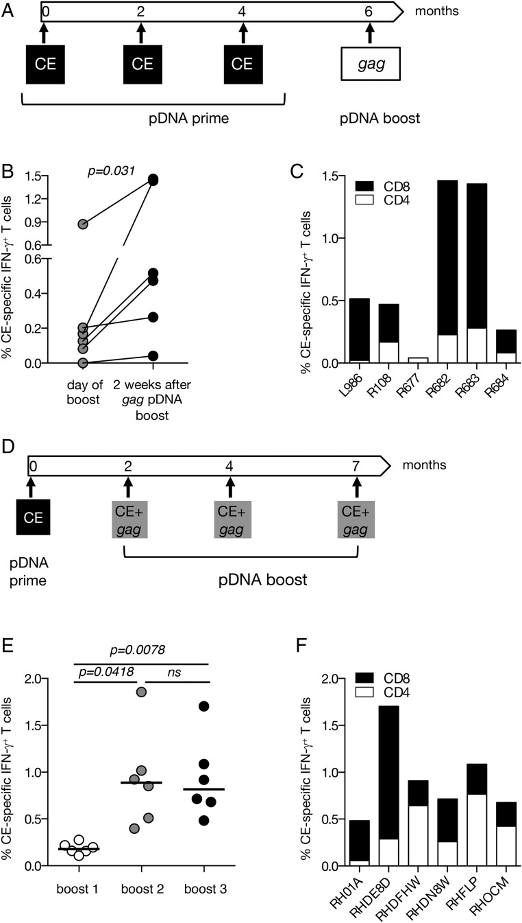
SIV p27CE pDNA prime-boost vaccination regimens. (A) The cartoon depicts the p27CE pDNA prime/gag pDNA boost vaccination regimen. (B) Plot shows the comparison between the levels of the CE-specific responses measured at the day of the gag pDNA booster vaccination and 2 wk later in six macaques described in Fig. 4. The p value is from a paired t test. (C) The frequency of p27CE-specific IFN-γ+ T cell responses is shown for CD4+ (open bars) and CD8+ (filled bars) T cells after the gag pDNA booster vaccination. (D) The cartoon depicts the p27CE pDNA prime and p27CE+gag pDNA booster vaccination regimen. (E) Comparison of the CE-specific cellular responses measured 2 wk after each p27CE+gag pDNA booster vaccination. The p values are from ANOVA (Dunnett’s test). (F) CE-specific T cell responses in the vaccinated macaques (n = 6) were measured 2 wk after the last p27CE+gag pDNA booster vaccination. The frequency of the IFN-γ+ p27CE-specific responses is shown for CD4+ (open bars) and CD8+ (filled bars) T cells.

