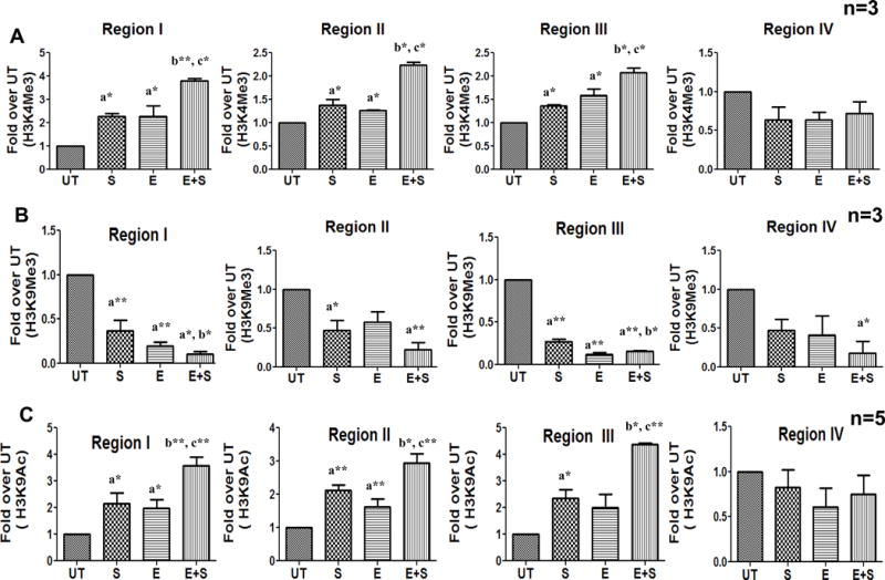
Figure 2. TCR stimulation-induced coordinate histone H3 modifications at the FasL promoter in control and ethanol treated CD4+ T lymphocytes.

Freshly isolated CD4+ T cells from healthy individuals (n=3–5) were examined by ChIP-qPCR analysis. The cells were untreated (UT) or exposed to 25mM ethanol (E) for 24h and subsequently stimulated with anti CD3/CD28 antibody (1μg/ml) for 6h (S and E+S). Histone modifications were assessed by analyzing chromatin that was immunoprecipitated with (A) anti-trimethylated Histone H3 lysine 4 (H3K4Me3) (B) anti-trimethylated Histone H3 lysine 9 (H3K9Me3) and (C) anti-acetylated Histone H3 lysine 9 (H3K9Ac) antibodies. Levels of histone modifications were measured using primer for regions I–IV shown in Figure 1. Differences are expressed as fold over UT after normalizing for input DNA. Results are represented as mean ± SE. Statistical analysis was performed by RMME models with Bonferroni’s correction for multiple comparisons. a* (p < 0.05) and a**(p < 0.01) compared to UT, b* (p < 0.05) and b**(p < 0.01) compared to S, and c* (p < 0.05) and c**(p < 0.01) compared to E.
