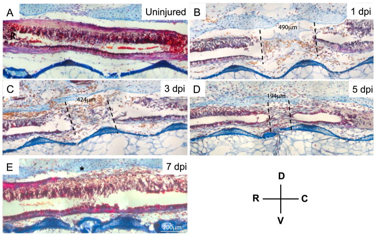
Fig. 1.
Spinal cord reconnection after spinal cord injury. Histological, Acid fucidin orange green (AfoG) staining of longitudinal sections of uninjured (A) versus injured spinal cords (B–E). At 1 day post injury an injury site of 490 μm is visible between the rostral and caudal ends of the severed spinal cord. Over time the distance between the rostral and caudal ends of the spinal cord decreases and the severed ends seal over forming terminal vesicle like structures (C and D). By 7 days post injury in animals that are 3–5 cm long the rostral and caudal ends of the spinal cord have reconnected and the central canal is reconnected (E). The location of the terminal vesicle in the rostral and caudal spinal cord is denoted by the dotted lines. The distance (micrometers) between the rostral and caudal terminal vesicles throughout regeneration is shown between the dotted lines. * denotes the original injury site. Each time point N=10.

