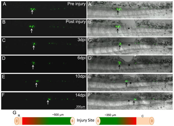
Fig. 2.
Ependymoglial cells from both the rostral and caudal sides of the injury contribute to replacing the injured spinal cord. Ependymoglial cells were labeled using a GFAP promoter driving GFP (A and A′). Ablation injury was performed adjacent to the injury site, asterisk denotes the injury site and arrow marks cells that were followed (B and B′). Within 3 day post injury some of the labeled cells adjacent to the injury site died (arrow C and C′). 6 days post injury the labeled cells adjacent to the injury site increase in number (D and D′). At 10 days post injury the labeled cells are found directly within the regenerating lesion area (E and E′). By 14 days post injury, when regeneration of the lesion is almost complete, some cells that originated on the rostral side of the injury are now found caudal to the injury site (F and F′). Panel G is a schematic diagram to summarize all in vivo imaging results, it was found that cells within 500 μm on the rostral side and 350 μm on the caudal side of the injury would migrate and contribute to regeneration of the lesion. Cells that were labeled outside of these regions did not migrate and contribute to the regeneration of the missing tissue. Cells labeled rostral within 500 μm N=25, caudal within 500 μm N=23. Cells labeled 500 μm–1 mm away from injury, rostral N=16, caudal N=17. In each animal multiple cells were labeled and followed.

