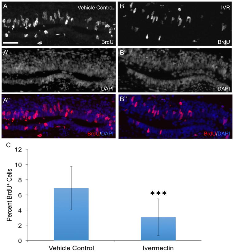
Fig. 4.
Prolonged depolarization inhibits ependymoglial cell proliferation in response to spinal cord injury. Animals were injected with vehicle control of PBS (n=15) (A, A′, A″) or ivermectin (IVR) to induce prolonged depolarization of the membrane (n=16) (B, B′, B″) prior to spinal cord ablation. One day after injury animals were subjected to an intraperitoneal injection with BrdU and harvested for staining 24 h later. (A″, B″) Comparison of the percent of BrdU+ cells in IVR treated and control axolotls shows there are significantly fewer BrdU+ cells in IVR treated animals 48 hours post injury compared to control axolotls (C). ***; P≤0.001. Error bars represent ± SEM. Scale bar is 75μm.

