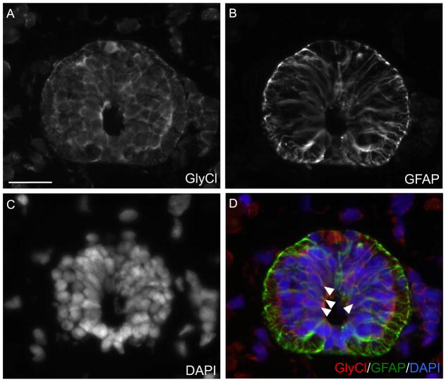
Fig. 5.
GlyCl-R is expressed by ependymoglial cells in the axolotl spinal cord. Cross sections of axolotl tails were stained with antibodies against (A) GlyCl-R or (B) the ependmyoglial marker GFAP. (D) Overlay with DAPI (Blue) GlyCl-R (red) is expressed by GFAP+ (green) ependymoglial cells (white arrows). Scale bar 50 μm. (For interpretation of the references to color in this figure legend, the reader is referred to the web version of this article.)

