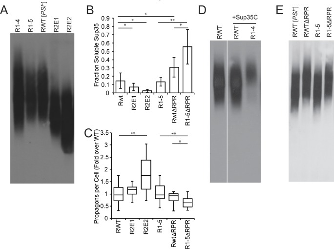
Fig 3. Sup35 repeats and RPR promote amyloid fragmentation.
A. SDD-AGE was performed on R1-4 (SY2057), R1-5 (SY2022), wildtype [PSI+] (SLL2606), R2E1 (SY2247), R2E2 (SY2300) lysates, followed by immunoblotting for Sup35. B. Cell lysates from wildtype [PSI+] (SLL2606), R2E1 (SY2247), R2E2 (SY2300), R1-5 (SY2022), ΔRPR (SY2023), and R1-5ΔRPR (SY1633) were incubated at 53°C and 100°C in the presence of SDS before SDS-PAGE and immunoblotting for Sup35. The amount of soluble Sup35 was determined as the percentage of signal at 53°C relative to 100°C. Bars represent means, error bars represent standard deviations. n≥5, *p<0.05, **p<0.01, student’s t-test. C. The number of propagons in wildtype [PSI+] (SLL2606), R2E1 (SY2247), R2E2 (SY2300), R1-5 (SY2022), ΔRPR (SY2023), and R1-5ΔRPR (SY1633) strains was determined by an in vivo colony based dilution assay. Horizontal lines on boxes represent the 25th, 50th, and 75th percentiles. Whiskers represent maximum and minimums. n≥13, *p<0.05, **p<0.01, student’s t-test. D. SDD-AGE was performed on wildtype [PSI+] (SLL2606), wildtype [PSI+] expressing an extra copy of Sup35C (SY2466) and R1-4 expressing an extra copy of Sup35C (SY2467) lysates, followed by immunoblotting for Sup35. Panels represent non-consecutive lanes run on the same gel. E. SDD-AGE was performed on wildtype [PSI+] (SLL2606), ΔRPR (SY2023), R1-5 (SY2022), and R1-5ΔRPR (SY1633) lysates, followed by immunoblotting for Sup35.

