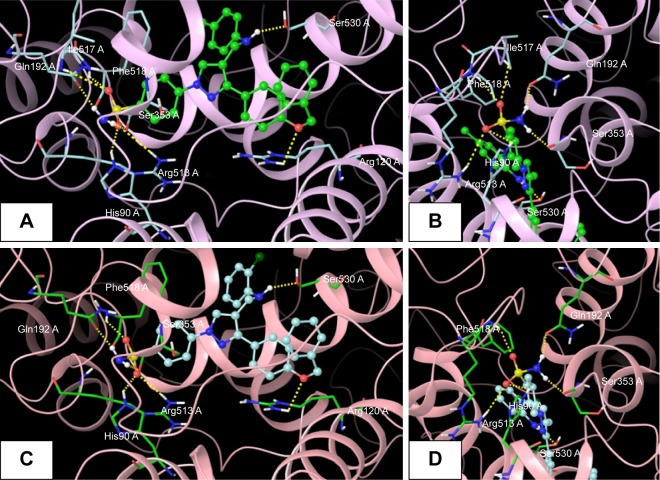
Figure 6.
Docked pose of 5s and 5u.
Notes: (A) Best docked pose of compound 5u (green), represented as ball-and-stick model in the binding site of COX2, showing hydrogen-bond interaction (yellow dashed lines) with Ser530, Arg120, His90, Arg513, Phe518, Ser353, Gln192, and Ile517; (B) zooming in on the docked-pose sulfonamide structure of compound 5u (green), showing hydrogen-bond interaction; (C) best docked pose of compound 5s (turquoise), represented as ball-and-stick model in the binding site of COX2, showing hydrogen-bond interaction (yellow dashed lines) with Ser530, Arg120, His90, Arg513, Phe518, Ser353, and Gln192; (D) zooming in on the docked-pose sulfonamide structure of compound 5s (turquoise), showing hydrogen-bond interaction.

