Figure 3.
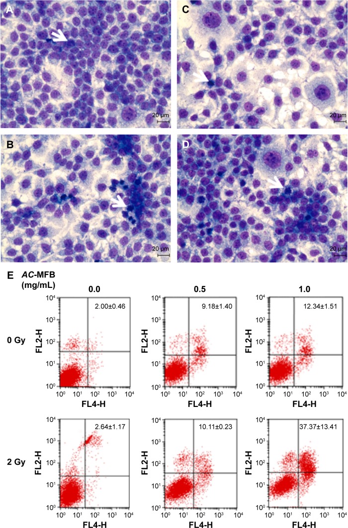
AC-MFB induced mitotic arrest morphology change and apoptosis of CE81T/VGH cell.
Notes: Cells were not treated (A, control) or treated with 0.5 and 1.0 mg/mL AC-MFB for 24 h (B), irradiation of 2 Gy (C), or AC-MFB plus irradiation (D). Arrows indicate representative cells characteristic of mitotic arrest. (E) Cellular apoptotic percentages induced by AC-MFB. Cell image was obtained by Liu’s Stain and microscopy at magnification ×400. Cell apoptosis percentages were determined by flow cytometry with Annexin-V/PI staining. Data are expressed as mean ± SD for three independent experiments.
Abbreviations: AC-MFB, Antrodia cinnamomea mycelial fermentation broth; Gy, gray; h, hours; PI, propidium iodine; SD, standard deviation.

