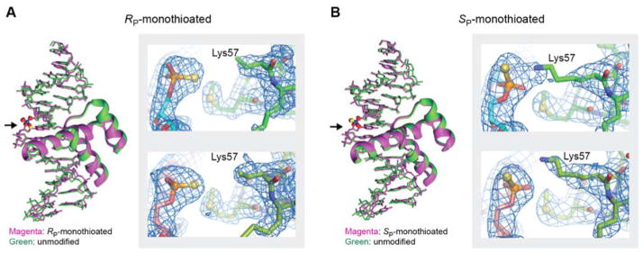
Figure 2.
Crystal structures of the Antp homeodomain-DNA complexes containing RP phosphoromonothioate (Panel A) or SP phosphoromonothioate (Panel B) at the Lys57 interaction site. The crystal structures of the RP and SP-monothioated DNA complexes were determined at 2.09 and 2.75 Å resolutions, respectively. For each panel, superposition of the crystal structures of the modified (green) and unmodified (purple) complexes is shown. The modification sites are shown in ball-stick representation. (indicated by arrows). For each modified complex, the electectron density map, of the phosphoromonothioate at Lys57 interaction site, for two structures in the asymmetric unit is shown. For the RP-monothioated DNA complex, the tip (i.e., Nξ, Cε, and Cδ groups) of the Lys57 side chain was not resolved in the crystallographic electron density map.

