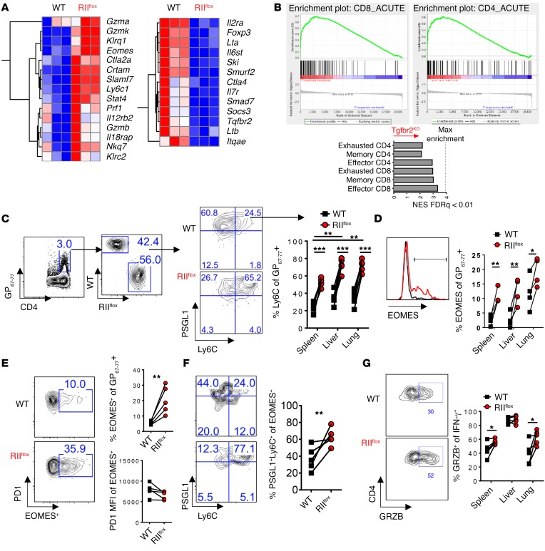
Figure 2. TGFβ-RII signaling in CD4 T cells suppressed terminal differentiation and the cytotoxic gene program early after chronic LCMV infection.
Reconstituted 1:1 mix of WT (CD45.1, black) and ERCre+ Tgfbr2fl/fl (RIIflox-CD45.2, red) bone marrow–chimeric mice were tamoxifen treated, rested, and infected with 2 × 10⁶ PFU of LCMV Cl13 and spleens, livers, and lungs were analyzed after 9 days. (A) FACS-sorted CD4+CD8–PD1+CD49d+ cells from each compartment were analyzed by microarray. Representative genes shown as a heat map of relative expression values (blue = min; red = max) from differentially regulated genes (P < 10–7). (B) GSEA and normalized enrichment score (NES) of TGFβ-RII CD4 T cell array signature for virus-specific CD4 and CD8 T cell microarrays from acute (effector and memory) and chronic (exhausted) LCMV infection (FDR q < 0.01). (C) PSGL1 vs. Ly6C gated on CD4 I-Ab:GP67–77 in the indicated tissue. (D) EOMES expression gated on PSGL1+Ly6C+ cells from C. (E) EOMES and PD1 expression on virus-specific CD4 T cells. (F) PSGL1 and Ly6C on cells from E. (G) Granzyme B expression in IFN-γ+ cells stimulated with cognate GP67–77 peptide from (Figure 1F). Representative of 3 independent experiments, with n = 4 or 5 mice/experiment. Paired t test, *P < 0.05, **P < 0.005, ***P < 0.0005.

