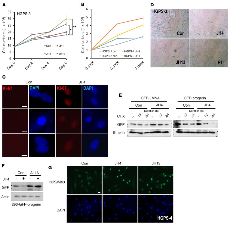
Figure 4. Antisenescence effect of the JH chemicals on HGPS cells.
(A) JH chemical overcame the growth suppression of HGPS cells. After seeding at 1 × 10⁴ cells per well, cell proliferation was determined by cell counting every 48 hours for 6 days. The experiment was performed in triplicate. *P = 0.01, **P = 0.016. P values were determined by Student’s t test. (B) Cell proliferation assay of normal cells obtained from a 9-year-old person (N9) under the same condition as above. JH4 could induce proliferation of normal cells, particularly at late time points (between 4 and 6 days). (C) Induction of Ki-67 by JH4 in HGPS cells. Three types of HGPS cells were incubated with JH4 for 48 hours and stained with Ki-67 Ab (red). Scale bars: 10 μm. (D) JH chemicals suppressed SA–β-gal expression in HGPS cells. SA–β-gal assay of HGPS cells incubated with JH chemicals (5 μM, for 48 hours). The same result was obtained for other HGPS cells (Supplemental Figure 7A). Original magnification, ×40. (E) JH4 suppressed the half-life of progerin. Pulse-chase analysis using the de novo synthesis inhibitor cycloheximide (CHX) showed a rapid reduction of progerin by treatment with JH4. Cells, transfected with GFP-LMNA or GFP-progerin vectors, were incubated with 10 μM CHX for the indicated durations, with or without 5 μM JH4. (F) The proteasome inhibitor ALLN blocked progerin downregulation by JH4. (G) JH chemicals induced H3K9Me3 in HGPS cells. Cells, incubated with the indicated chemicals (5 μM) for 48 hours, were fixed with 100% Me-OH and stained with anti-H3K9Me9 Ab (green). DAPI was used for DNA staining (blue). Scale bars: 10 μm. Con, control.

