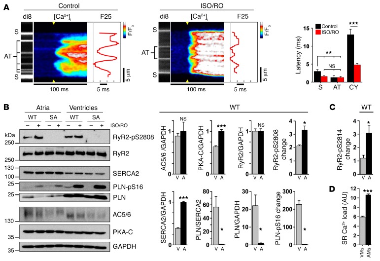
Figure 6. Catecholaminergic stimulation accelerates atrial Ca2+ signaling.
(A) Live imaging of AT and S membrane structures during Ca2+ transient onset in control- versus ISO/RO-treated AMs (0.5-Hz pacing). ISO/RO treatment accelerated transversal Ca2+ release propagation across the cytosol, compressing the F25 profile through a decreased cytosolic (CY) latency of early Ca2+ release (dF/dt). Data are representative of 9 control- and 17 ISO/RO-treated AMs. **P < 0.01 and ***P < 0.001, by Student’s t test. (B) Immunoblots comparing ISO/RO-treated (1 μM/10 μM) atrial (A) tissue from WT and phosphorylation-incompetent RyR2-S2808A+/+ mouse hearts; ventricular (V) tissue was compared for reference. In WT atria, significantly higher expression levels of both PKA-C and SERCA2 were detected, in contrast to significantly lower PLN levels. Atrial versus ventricular PKA phosphorylation changes were significantly increased for the RyR2-S2808 site, but not for the PLN-S16 site. Note the similar AC5/6 and RyR2 protein levels. Blots are representative of 3 individual experiments (see Supplemental Figure 10 for uncut blots). (C) Analogous RyR2-S2814–specific immunoblotting established in CaMK phosphorylation–incompetent RyR2-S2814A+/+ mouse hearts (see also Supplemental Figures 12A and 12B). Bar graph shows a significantly increased atrial RyR2-S2814 phosphorylation change in atrial tissue after ISO/RO (1 μM/10 μM) treatment. (D) SR Ca2+ load was significantly higher in AMs than in VMs measured with caffeine (10 mM). n = 8 AMs, 9 VMs. *P < 0.05 and ***P < 0.001, by Student’s t test.

