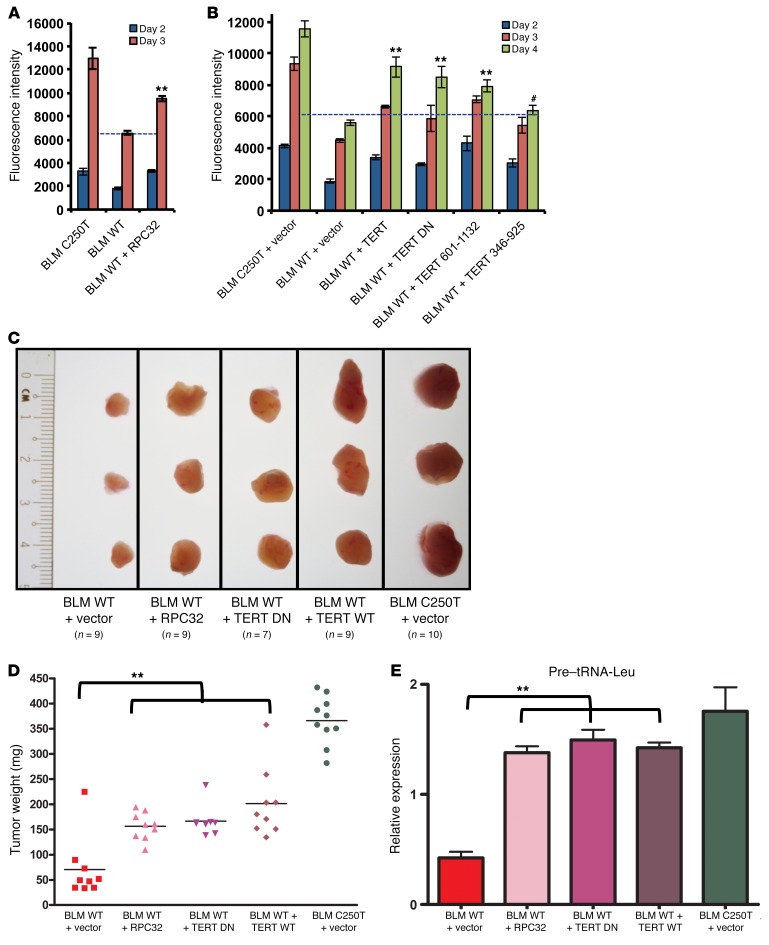
Figure 7. RPC32 and TERT can rescue cell proliferation upon TERT depletion.
(A) A cell proliferation assay was performed in BLM C250T, BLM WT, and BLM WT cells infected with RPC32-expressing vector. Graph shows fluorescence intensity measured using an Alamar Blue viability assay. n = 3. (B) A cell proliferation assay was performed in BLM C250T, BLM WT, and BLM WT cells infected with TERT or TERT DN or with TERT 601-1132 or TERT 346-925 constructs. n = 3. (C) BLM WT cells were infected with vector or TERT or with TERT DN or RPC32 and expanded along with BLM C250T cells infected with vector. Following infection, the cells were xenografted s.c. into NOD/SCID mice and allowed to form tumors. After 15 days, tumors were harvested and analyzed. Figure shows images of 3 independent tumors of each cell type; the number of tumors obtained is indicated. (D) Weights of tumors produced in C. (E) RNA was extracted from tumors obtained from C. Graph shows the relative expression of pre–tRNA-Leu normalized against actin levels in tumors. All error bars indicate the mean ± SEM. **P < 0.001 and #P > 0.05, by 1-way ANOVA with Tukey’s multiple comparisons test.

