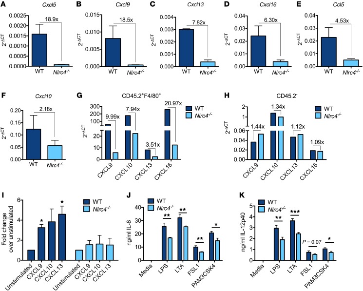
Figure 5. Absence of NLRC4 in macrophages alters the tumor cytokine and chemokine milieu.
(A–F) WT and Nlrc4–/– mice were injected s.c. with 1 × 10⁵ B16F10 cells. On day 12 after inoculation, total RNA was isolated from homogenized tumors and used to determine cytokine and chemokine expression via quantitative qPCR utilizing a PCR array. Selected genes from the array are displayed; data are pooled from 3 separate experiments (n = 3 mice per group). (G and H) WT and Nlrc4–/– mice were injected s.c. with 1 × 10⁵ B16F10 cells; 14 days after inoculation, tumors were harvested, pooled, and FACS sorted based on CD45.2 and F4/80 staining. RNA was isolated from CD45.2- and CD45.2+F4/80+ cells and used to determine Cxcl9, Cxcl10, Cxcl13, and Cxcl16 expression by qPCR; data are representative of 2 independent experiments with n ≥ 5 pooled tumors per group. (I) WT and Nlrc4–/– BMDMs were challenged for 9 hours with B16F10 whole tumor homogenate. Cxcl9, Cxcl10, and Cxcl13 expression was determined by qPCR. Data are pooled from 3 independent experiments, and fold change in gene expression is relative to unstimulated samples. (J and K) WT and Nlrc4–/– BMDMs were challenged with 50 ng/ml LPS, 50 μg/ml LTA, 100 ng/ml FSL-1, and 1 μg/ml Pam3CSK4. Twenty hours later, supernatants were collected and levels of IL-6 (J) and IL-12p40 (K) determined by ELISA; data are representative of 3 independent experiments. (A–F and I) Error bars represent SEM. (J and K) Error bars represent SD. (I–K) *P ≤ 0.05, **P ≤ 0.01, and ***P ≤ 0.001, unpaired 2-tailed Student’s t test.

