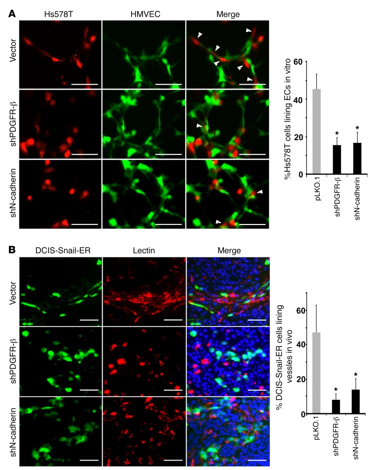
Figure 5. PDGFR-β and N-cadherin are required for EMT cancer cells to associate with endothelial cells in vitro and in vivo.
(A) Control, PDGFR-β–, or N-cadherin–depleted Hs578T cells (red) were cocultured with HMVECs (green) for tube formation in vitro. EC-bound Hs578T cells are denoted by white arrowheads and quantified (right). (B) Control, PDGFR-β–, or N-cadherin-depleted DCIS-Snail-ER cells (green) were mixed with unlabeled DCIS cells (at a 1:4 ratio) for orthotopic implantation in mice. All tumor-bearing mice received tamoxifen treatment. Tumor sections were stained with Griffonia simplicifolia lectin for vasculature (red). DNA was stained blue with Hoechst 33342. DCIS-Snail-ER cells aligned with vasculature are quantified (right). Scale bars: 50 μm. Error bars represent SD (n = 4–6). *P < 0.05. Statistical differences were determined by 2-tailed Student’s t test.

