
Fig. 1.
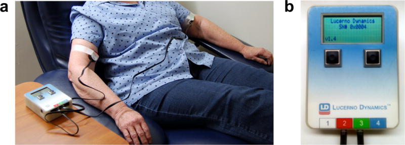
(a) Sensors containing scintillation material convert gamma radiation, emitted from the decay of 18F-FDG, to visible light in the form of photon counts/sec. For recordings of the injection arm and control arm, sensors are positioned over the bicep, since the convex shape of the antecubital fossa hinders direct recording from this region. Sensors are held in place using standard medical tape. (b) Sensors are connected via cable to a portable reader module that actively records activity counts at 1-second intervals. Data from each sensor channel (up to four) are digitized, tabulated and exported from the reader in a variety of formats for further computational analysis.