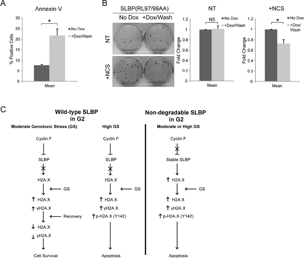
Figure 6. SLBP(RL97/99AA) expression in G2 leads to increased apoptosis upon genotoxic stress.
(a) U2OS cells prepared as in Figure 5A were collected 4 hours after treatment with NCS, stained with Annexin-V Alexa-488 conjugate and propidium iodide, and analyzed by flow cytometry. Data is presented as the percent of cells that stained positive for Annexin-V and negative for propidium iodide (*p≤0.05, n=2).
(b) Non-treated (NT) U2OS or U2OS cells treated with NCS (+NCS) were prepared as in Figure 5A, and allowed to grow for approximately 12 days, until colonies could be identified. Each plate was then fixed with 6% glutaraldehyde and stained with crystal violet. The data are presented as mean ± SD (*p≤0.05, NS; not significant, n=3, each in triplicate).
(c) A model of the regulation of survival and apoptosis by the cyclin F-SLBP axis. During G2, after the large majority of DNA replication has occurred, cyclin F accumulates, thereby promoting SLBP degradation. Cells in G2 are able to survive moderate genotoxic stress, whereas high levels of genotoxic stress leads to apoptosis. Stabilization of SLBP into G2 induces high expression of H2A.X and sensitizes the cell to genotoxic stress.

