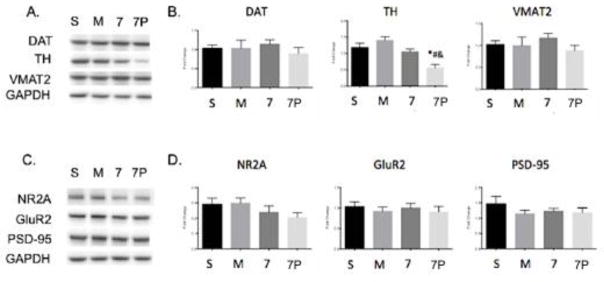
Figure 8.
Whole brain protein levels of METH neurotoxicity markers after treatment with saline, METH, scFv7F9Cys, or scFv7F9Cys-PEG20K. A. Representative Western blots of dopaminergic markers: DAT, TH, and VMAT2. B. Quantification of Western blots for DAT, TH, and VMAT2. C. Representative Western blots of glutamatergic markers: NR2A, GluR2, PSD-95. D. Quantification of Western blots for NR2A, GluR2, and PSD-95. Error bars are SEM. S – Saline, M – METH, 7 – scFv7F9Cys, 7P – scFv7F9Cys-PEG20K, * - compared to saline (p<0.05), # - compared to METH (p<0.05), & - compared to scFv7F9Cys (p<0.05)

