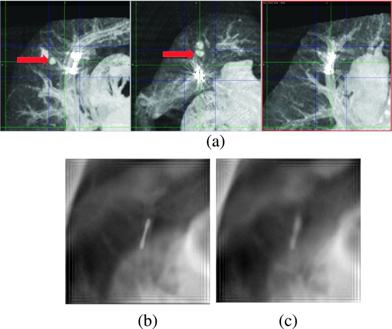
FIG. 7.
(a) Sagittal, axial, and coronal MIPs of the lungs, respectively. The dark (blue) lines show the edges of the MIPs in the other two orientations. The arrows show the lesion. (b) The SBDX image of the plane of the fiducial. (c) The SBDX image of the plane of the lesion. Unlike the coronal MIP, the lesion is not visible in the SBDX images. There is not sufficient blurring of the fiducial to resolve the lesion.

