Fig. 6.

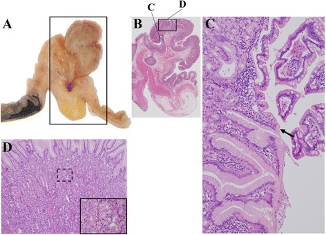
Histological appearance of the polypoid lesion specimen (a, b) showing a transition between the ectopic gastric mucosa and the intestinal mucosa (c, arrow). Ectopic gastric mucosa of the fundic type (d)

Histological appearance of the polypoid lesion specimen (a, b) showing a transition between the ectopic gastric mucosa and the intestinal mucosa (c, arrow). Ectopic gastric mucosa of the fundic type (d)