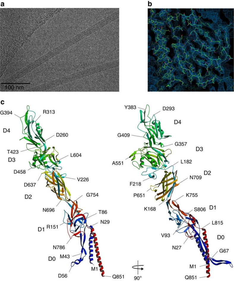
Figure 1. Complete structure of FlgE from C. jejuni.
(a) Cryo-electron micrograph of FlgEcj. (b) Cryo-EM density map superimposed with part of the structure of FlgEcj. (c) Two different views of the Cα backbone tracing of FlgEcj with its five domains, D0, D1, D2 D3 and D4. The chain is colour coded in a spectral (‘rainbow') sequence, from blue at the N-terminus through red at the C-terminus. Figure prepared Figure prepared with Coot36, MOLSCRIPT44 and RASTER3D (ref. 45).

