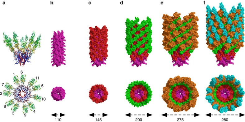
Figure 2. Domain distribution in the hook of C. jejuni.
(a) Lateral and top views of two turns of the hook of C. jejuni showing the eleven molecules that mark the start of the eleven protofilaments of the hook. (b–f) Lateral and top views of the hook of C. jejuni showing the organization and the packing of the different layers made by the domains of FlgE. Domain D0 in purple, domain D1 in red, domain D2 in green, domain D3 in brown and domain D4 in cyan. The image at left shows only D0. Additional domains are added in each subsequent image. The approximate diameters are indicated at the bottom. Figures were prepared with PyMOL (The PyMOL Molecular Graphic System, Schrödinger, LLC. http://www.pymol.org.).

