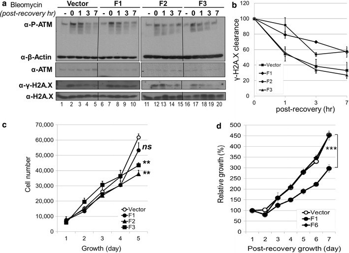
Fig. 4.

Bleomycin susceptibility assays for the ectopic expression of ATM-interacting p400 fragments. a Time course of DNA damage response after bleomycin exposure. Puromycin-selected U2OS cells were exposed to bleomycin at 10 μg/ml for 1 h, replaced with normal growth medium and incubated further for an indicated period as a post-recovery time. Whole cell extracts and histone extracts were analysed by immunoblotting with antibodies as indicated. b γ-H2A.X clearance after bleomycin exposure. The signals of γ-H2A.X to total H2A.X on the immunoblot in (a) were quantified in triplicate with Image J software. The peaked ratio right after the bleomycin treatment (0 h post-recovery) set to 100 and the relative changes were calculated. c Growth curve of puromycin-selected U2OS cells. U2OS cells were infected with lentivirus expressing Flag-F1, Flag-F2 or Flag-F3, and selected for 5 days using puromycin. Each cell line was seeded in 96-well plate in triplicate and monitored for the cell proliferation. (Growth day 5: n = 3, *p < 0.05, **p < 0.01, ns: not significant by the Student t test compared to the vector). The experiments were repeated triple times with similar results. d Bleomycin sensitivity assay of U2OS cells. U2OS cells stably expressing Flag-F1 or Flag-F6, were exposed to bleomycin at 10 μg/ml for 12 h and seeded in 96-well plate in triplicate to assess the bleomycin sensitivity. The cell number observed at the first day of post-recovery set to 100% and the relative cell growth was calculated during the post-recovery period. (Post-recovery day 7: n = 3, ***p < 0.001)
