
Figure 1.
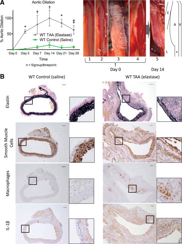
A, Aortic dilation over time for wild-type (WT) mice exposed to elastase (gray) and saline (green). *P<0.001, †P<0.0001, ‡P<0.01. Sample photographs of the thoracic aortic aneurysm (TAA) model showing (1) initial exposure of the thoracic aorta through a left thoracotomy, (2) dissection of the pleura, (3) application of an elastase soaked sponge, and (4) sponge removal. For aortic harvest (5), the thoracotomy was reopened, and thoracic aorta was dissected free and measured. Illustration to the right demonstrates relative association of aorta (A) with esophagus (E), lung (L), and hemiazygos vein (V). The internal control section of unexposed aorta is marked with *. B, Sample cross-sections of WT control and WT TAAs on day 14 staining for elastin, smooth muscle cells, macrophages, and interleukin-1β (IL-1β). Scale bar, 50 μm.