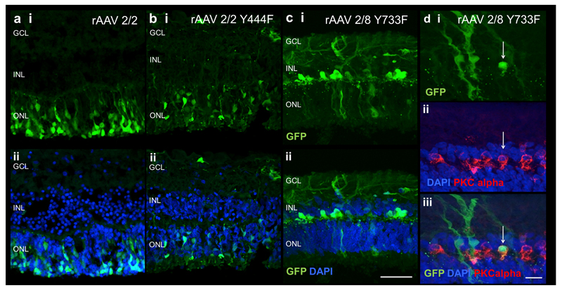
Figure 6.
GFP fluorescence following ex vivo administration of AAV vectors in human retinal tissue. GFP expression in explants following rAAV 2/2 (a), rAAV2/2 Y44F (b) and rAAV2/8 Y733F (c) exposure. Histological cross-sections of specimens show expression of GFP (green, panel i) and merge with DAPI (blue, panel ii) for each vector. Images d shows colocalisation of GFP (green) in bipolar cells identified by PKC alpha immunostaining (red) demonstrating human bipolar cell transduction. GCL= ganglion cell layer; INL= inner nuclear layer, ONL= outer nuclear layer. Scale bar 50μm for images a-c, 10μm for images d.

