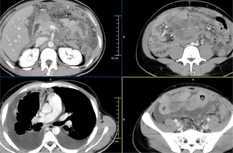
Fig. 4.
MDCT transverse sections shows extensive peritoneal, omental lesions, and ascites.Large confluent adenopathy is seen in the retro peritoneum encasing the aorta, celiac axis ,superior mesenteric artery and their branches.CT sections of the Chest showed bilateral pleural collections. Patient was subsequently diagnosed to have high grade Non-Hodgkin’s Lymphoma on lymph node biopsy

