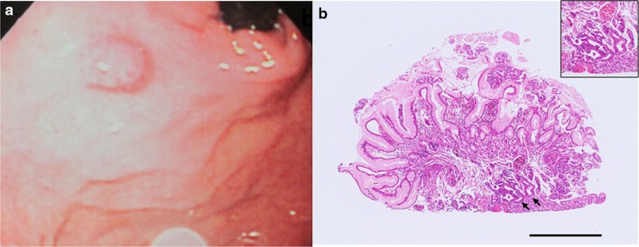
Fig. 1.
Endoscopic appearance of the lesion and histologic features of the biopsy specimen obtained upon the initial endoscopic examination (H&E stain). a The initial endoscopic examination revealed a reddish, 10-mm, elevated lesion on the posterior wall of the fornix (shown here in the polaroid photograph obtained at the time). b Non-neoplastic glands had been detected in the initial biopsy specimen, but upon re-examination of the original specimen after endoscopic treatment, we noted atypical glands corresponding to GAFG (black arrows). Black bar 1 mm. Inset high magnification view of the GAFG

