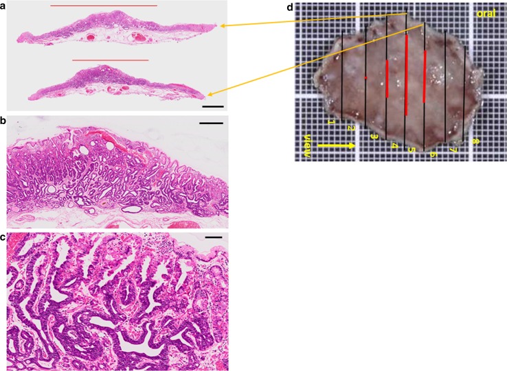
Fig. 4.
Histopathologic features of the ESD specimen. a Histologic slides under loupe magnification showing the spread of the adenocarcinoma. Black bar 2 mm. b Low magnification image shows irregularly shaped neoplastic glands lying mainly in the lower portion of the oxyntic mucosa. Black bar 500 μm. c High magnification image. Note the irregularly shaped glands consisting of two types of atypical glandular cells, those with basophilic cytoplasm, and those with eosinophilic cytoplasm. Black bar 200 μm. d Spread of the carcinoma mapped on the surgical specimen. Black line cut line, Red line spread of the carcinoma. Orange arrows from the cut lines point to the respective loupe images (a)

