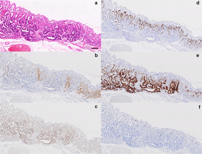
Fig. 5.
Histologic and immunohistochemical features of the adenocarcinoma and non-neoplastic fundic glands. a H&E stained tissue. The border between GAFG and non-neoplastic oxyntic mucosa is evident. b Pepsinogen-I staining. The neoplastic glands are more weakly stained than the non-neoplastic fundic glands. c Staining for MIST-1. Many positively stained cells are evident in the neoplastic glands. d Staining for H+/K+-ATPase. Positively stained neoplastic cells are prominent in the upper portion of the mucosa. e Staining for MUC6. The distribution of positively stained neoplastic cells corresponds well with that of cells stained positively for MIST-1. f Ki-67 staining. Black bar 200 μm

