
Fig. 6.
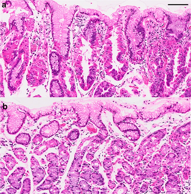
Histologic comparison of neoplastic glands and non-neoplastic fundic glands (hematoxylin and eosin-stained sections). a Protrusions are visible in the neoplastic cells showing not only parietal cell but also chief cell differentiation. b Protrusions are not seen in the non-neoplastic parietal cells. Black bar 100 μm