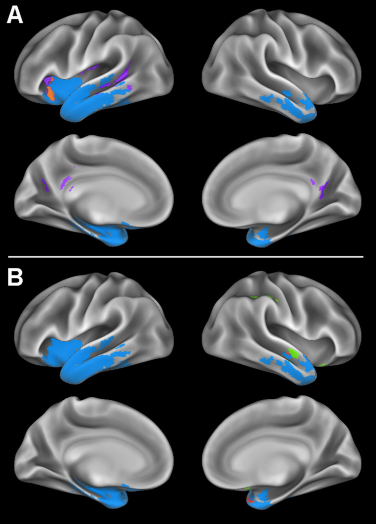
Figure 1. Whole-brain cortical thickness and perfusion comparisons.
A) Regions where patients with semantic variant primary progressive aphasia (svPPA) display gray matter thinning relative to controls are shown in blue, regions of partial volume corrected (PVC) hypoperfusion without gray matter thinning in svPPA are shown in purple, and a region displaying both PVC hypoperfusion and gray matter thinning relative to controls is shown in orange. B) Regions where patients with svPPA display gray matter thinning relative to controls are shown in blue, PVC hyperperfusion without gray matter thinning in svPPA is shown in green, and regions where patients display both PVC hyperperfusion and gray matter thinning relative to controls are shown in red.

