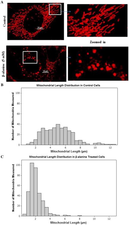
Fig. 3.

β-alanine mediates mitochondrial fragmentation. Control and β-alanine-treated cells were incubated for 48 h. a Shown are representative MitoTracker Deep Red (150 nM) loaded control fibroblasts (upper panel), which contain tubular mitochondria, and β-alanine-treated fibroblasts (lower panel), which contain fragmented mitochondria. b Average distribution of mitochondrial length of control fibroblasts in culture. c Average distribution of mitochondrial length of β-alanine-treated fibroblasts. The distribution pattern of mitochondrial length was shifted to the left by β-alanine treatment
