
Fig. 4.
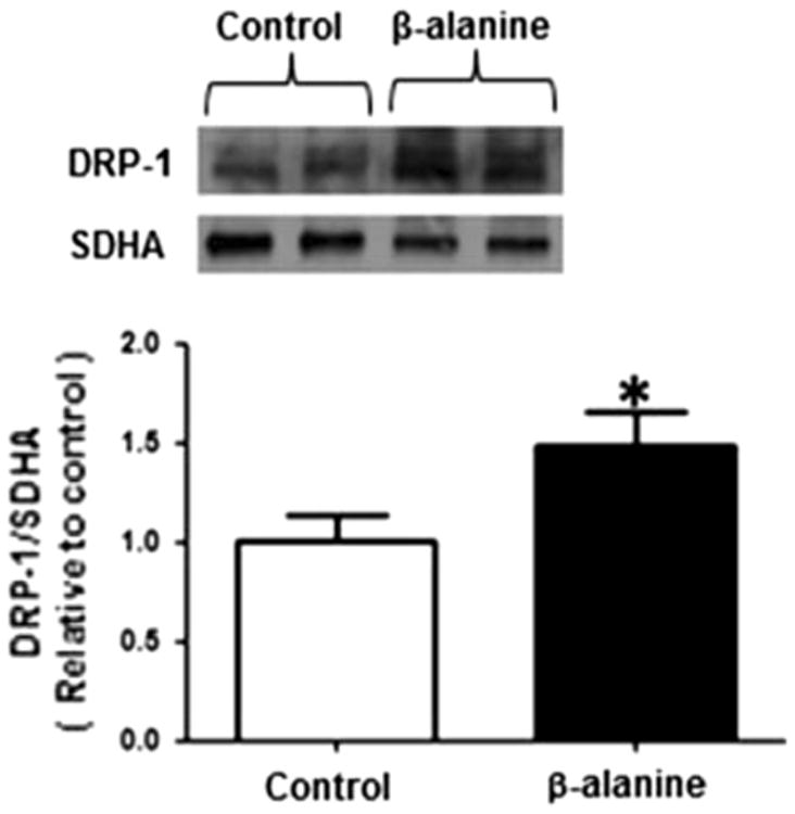
β-alanine treatment is associated with an increase in mitochondrial Drp1 content. Mitochondria were isolated from control and β-alanine-treated fibroblasts and then subjected to Western blot analysis of Drp1. The upper panel depicts a representative Western blot for mitochondrial Drp1 content of control and β-alanine-treated cells, with succinate dehydrogenase (SDHA) serving as a loading control. Data shown in the lower panel represent mean ± S.E.M. of eight different preparations. The asterisk denotes a significant difference in mitochondrial Drp1 content of control and β-alanine-treated cells (p < 0.05)