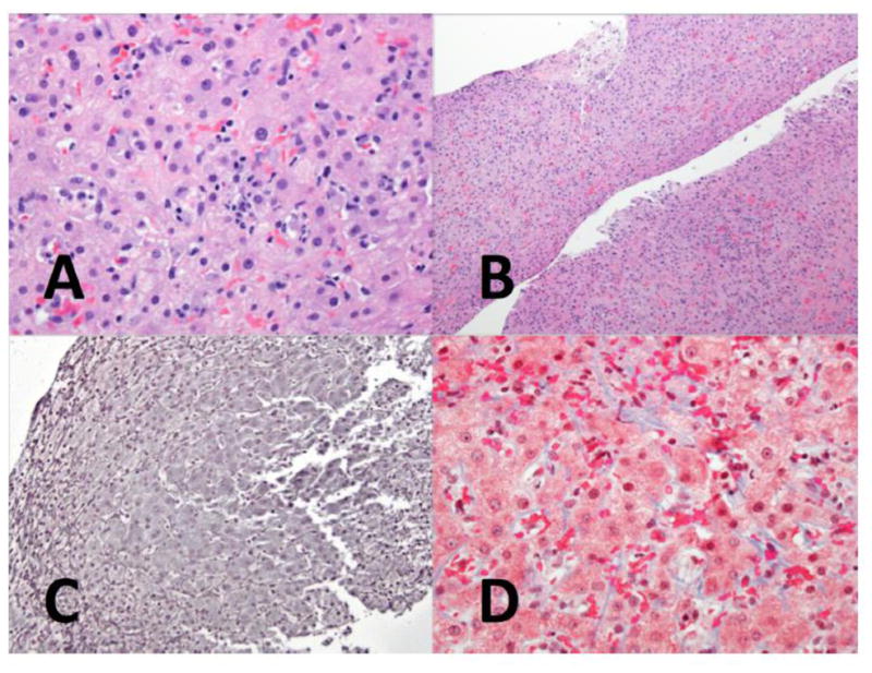
Figure 1.

A. Mild lobular inflammation is seen in the parenchyma (H&E, 400x)
B. At medium magnification, the hepatic parenchyma shows few abnormalities. (H&E, 100x)
C. Reticulin staining shows the characteristic changes of NRH, with widened, 2-cell thick liver plates bounded by compressed atrophic liver cell plates. (Reticulin, 200x)
D. A trichrome stain for collagen shows only mild perisinusoidal fibrosis (Masson trichrome, 400x)
