
Figure 2.
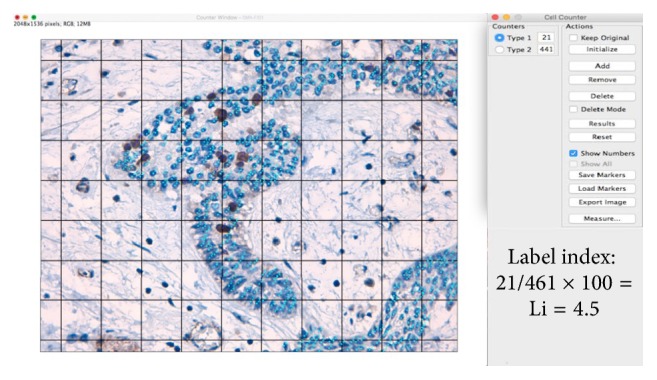
Photomicrograph of a solid ameloblastoma specimen at 400x magnification. This photomicrograph represents the manual cell counting method (DM scoring) using the Grid and Cell Counter plugins. The Grid was constructed with an area-per-point of 17,046 pixels∧2. The light blue dots indicate nuclei negative for DAB or Ki-67 staining. The dark blue nuclei indicate positive DAB and Ki-67 staining (ImageJ, method proposed by Carreón-Burciaga et al. [8]).