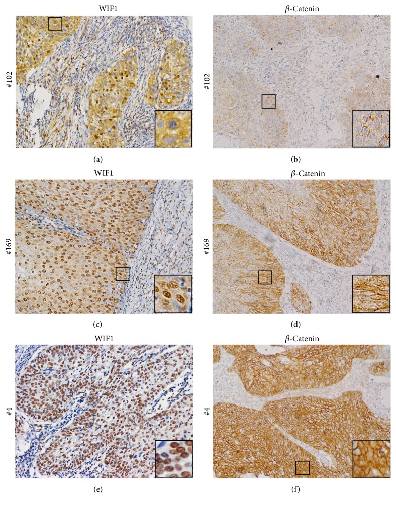
Figure 2.
Comparison of WIF1 and β-catenin expression in CC. The relationship of WIF1 and β-catenin immunostaining for three representative cases: ((a), (b)) Case 102, indicating strong WIF1 cytoplasmic staining and negative β-catenin cytoplasmic/nuclear staining; ((c), (d)) Case 169, which shows moderate immunostaining for both WIF1 and β-catenin; ((e), (f)) Case 4, showing negative WIF1 cytoplasmic staining and strong β-catenin cytoplasmic staining. Magnification: ×200. Insets are magnified images from selected areas (small squares).

