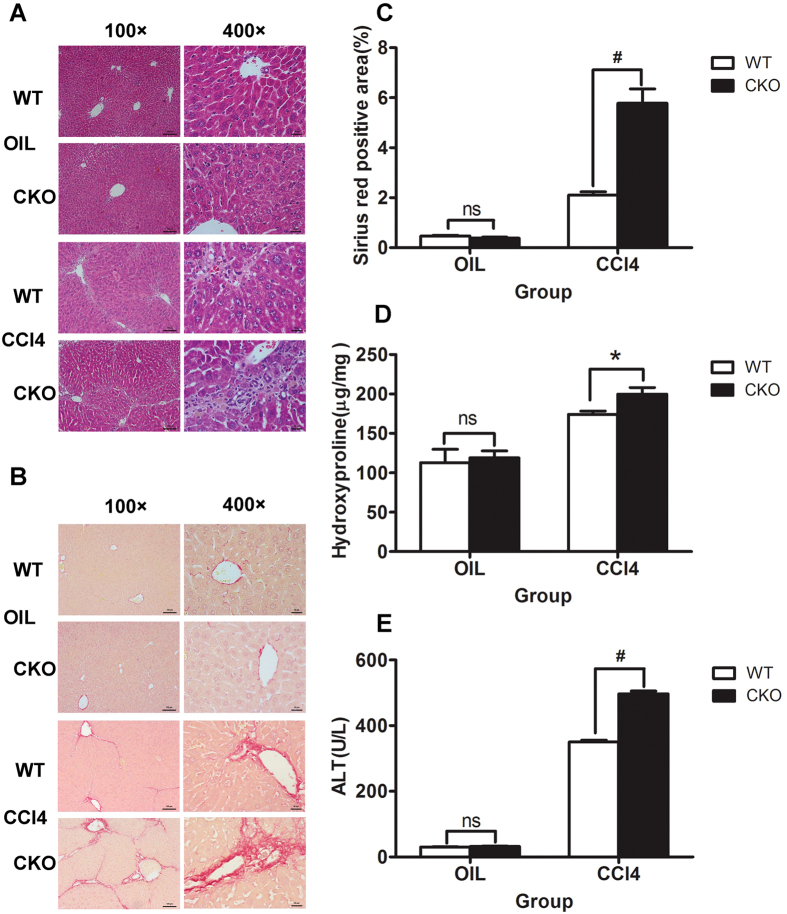
Figure 2. TSC1 deletion augments CCl4− induced liver fibrosis in mice.
Tamoxifen-treated control [wild-type (WT)] and TSC1 conditional knockout (CKO) mice received intraperitoneal injections of either CCl4 or oil, as indicated. H&E staining (A) and Sirius Red staining (B) were performed 8 weeks later. TSC1 CKO mice had more severe liver fibrosis than WT mice. All were imaged at 100x or 400x magnification. (C) The Sirius Red staining area in the liver was calculated by Image-Pro Plus 6.0. in six different images taken at 100x magnification on each slide. (D) The liver samples were analyzed for hydroxyproline content. (E) Liver injury was determined by serum ALT in WT and TSC1 CKO mice. The results are presented as the mean ± SEM derived from three independent experiments. NS, p > 0.05; *p < 0.05; #p < 0.01 for TSC1 CKO vs. WT mice.

