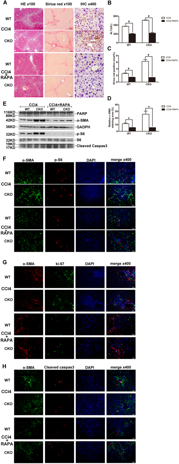
Figure 6. Rapamycin attenuates CCl4− Induced Liver Fibrosis and reverses phenotypes in TSC1 CKO mice.

(A) Liver tissue samples from CCl4− or CCl4+ rapamycin- treated mice for 8 weeks were analyzed for histological analysis. As shown in figure H&E staining and Sirius Red staining, CCl4+ rapamycin- treated mice had less severe liver fibrosis than CCl4− treated mice. All were imaged at 100x magnification. IHC staining was performed with α-SMA in CCl4− or CCl4+ rapamycin- treated mice liver tissues. (B) Liver injury was determined by serum ALT in CCl4− and CCl4+ papamycin-treated mice. (C) The Sirius Red staining area in the liver was calculated by Image-Pro Plus 6.0. in six different images taken at 100x magnification on each slide. (D) a-SMA staining area in the liver was calculated by Image-Pro Plus 6.0. in six different images taken at 400x magnification on each slide. (E) Liver tissue samples from CCl4− or CCl4+ rapamycin- treated mice were analyzed for PARP, cleaved caspase-3, p-S6, and S6 protein by western blot analysis. Immunofluorescence(IF) staining were performed to evaluate the impact of rapamycin on myofibroblasts proliferation and apoptosis. (F) Sections were stained with α-SMA (green) and p-S6 (red) antibodies. There were decreased α-SMA and pS6 double positive cells in mice administered CCl4+ rapamycin. (G) Sections were stained with α-SMA (red) and ki67 (green) antibodies.α-SMA- and ki67 double positive cells were decreased in mice following CCl4+ rapamycin treatment. (H) Sections were stained with α-SMA (green) and cleaved caspase-3 (red) antibodies. α-SMA- and cleaved caspase-3 double positive cells increased in the CCl4+ rapamycin-treated mice. Tissues were counterstained with DAPI (blue) to detect nuclei and imaged at 400x magnification. #p < 0.01; *p < 0.05 for CCl4− vs. CCl4+ rapamycin-treated mice.
