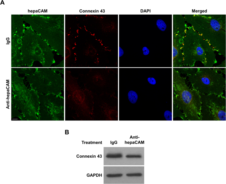
Figure 2. HepaCAM enhances connexin 43 expression.
(A) Treatment of hepaCAM-expressing U373 MG cells with antibodies against the hepaCAM extracellular domain prevents the association of hepaCAM with connexin 43 at cell-cell contacts. Wild-type hepaCAM-expressing U373 MG cells were treated overnight with antibody against the hepaCAM extracellular domain in soluble form (10 μg/ml). Cells were also treated with the isotype mouse IgG1 as a control. The next day, cells were fixed and immunofluorescent staining was performed with antibodies against the hepaCAM extracellular domain (green) and connexin 43 (red). Co-localization of hepaCAM and connexin 43 is indicated by yellow fluorescence. Nuclei were stained with DAPI (blue). Cells were visualized by confocal microscopy under a 60× objective. Scale bar: 10 μm. (B) Treatment of hepaCAM-expressing U373 MG cells with antibodies against the hepaCAM extracellular domain causes a downregulation of connexin 43 expression. Wild-type hepaCAM-expressing U373 MG cells were treated overnight with antibody against the hepaCAM extracellular domain in soluble form (10 μg/ml). Cells were also treated with the isotype mouse IgG1 as a control. The next day, cells were lysed and 20 μg of cell lysates were subjected to Western blot analysis using connexin 43 antibody. GAPDH was used as a loading control. The full view blots are shown in Supplementary Figure 2.

