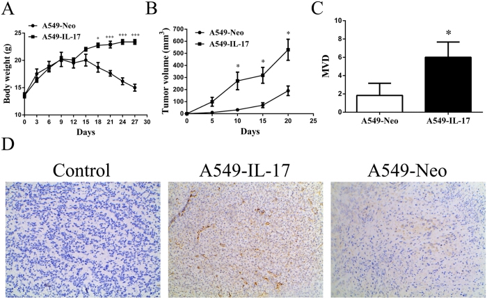
Figure 8. CD31 expression was increased in A549-IL-17 cell-bearing nude mouse tissues.
(A) The time course of in vivo growth of A549-IL-17 cells vs. A549-Neo cells in nude mice (n = 5). (B) The time course of increases in the tumour volumes in A549-IL-17 and A549-Neo cells in nude mice (n = 5). (C) The t-test was used to analyse the tumour microvessel densities in A549-IL-17 and A549-Neo cell-bearing nude mice (n = 5). (D) CD31 staining in tumour tissues from one each of A549-IL-17 and A549-Neo cell-bearing nude mice. For the control, one nude mouse tissue sample was not treated with a primary antibody. The data are presented as the mean ± SD for five mice per group, and the results are representative of two independent experiments. *p < 0.05; **p < 0.01; and ***p < 0.001.

