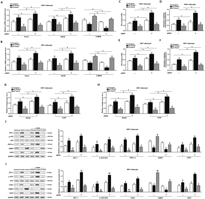
Figure 3. α-MSH and Foxc2 jointly increase fatty acid oxidation in white and brown adipocytes.
(A) mRNA levels of Foxc2, MC5R and C/EBPβ of iWAT adipocytes. adipocytes were pre-transfected with pc-Foxc2 and si-Foxc2 for 72 h and and then treated with 500 nM α-MSH for 1h before collected (n = 3); (B) mRNA levels of Foxc2, MC5R and C/EBPβ of iBAT adipocytes. Adipocytes were pre-transfected with pc-Foxc2 and si-Foxc2 for 72 h and then treated with 500 nM α-MSH for 1h before collected (n = 3); (C) FFA level of iWAT adipocytes with pc-Foxc2 or si-Foxc2 transfection for 72 h and 500 nM α-MSH incubation for 1 h (n = 3); (D) Palmitate oxidation of iWAT with pc-Foxc2 or si-Foxc2 transfection for 72 h and 500 nM α-MSH incubation for 1 h (n = 3); (E) FFA level of iBAT adipocytes with pc-Foxc2 or si-Foxc2 transfection for 72 h and 500 nM α-MSH incubation for 1 h (n = 3); (F) Palmitate oxidation of iBAT with pc-Foxc2 or si-Foxc2 transfection for 72 h and 500 nM α-MSH incubation for 1 h (n = 3); (G) Levels of MCAD and LCAD of iWAT adipocytes with pc-Foxc2 or si-Foxc2 transfection for 72 h and 500 nM α-MSH incubation for 1 h, protein level was detected by ELISA test (n = 3); (H) Levels of MCAD and LCAD of iBAT adipocytes with pc-Foxc2 or si-Foxc2 transfection for 72 h and 500 nM α-MSH incubation for 1 h, protein level was detected by ELISA test (n = 3); (I) Protein levels of CPT-1, p-ACC, PGC1-α, FABP4 and UCP2 in iWAT with pc-Foxc2 or si-Foxc2 transfection for 72 h and 500 nM α-MSH incubation for 1 h (n = 3); (J) Protein levels of CPT-1, p-ACC, Cidea, FABP3 and UCP1 in iWAT with pc-Foxc2 or si-Foxc2 transfection for 72 h and 500 nM α-MSH incubation for 1 h (n = 3). pc-Foxc2: adenovirus overexpression vector of Foxc2, si-Foxc2: lentiviral interference vector of Foxc2, CPT-1: carnitine palmitoyl transferase-1, ACC: acetyl-CoA carboxylase, ACC: acetyl-CoA carboxylase. Values are means ± SD. vs. control group, *p < 0.05.

