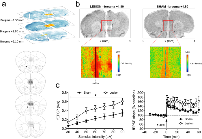
Figure 1. Acute hippocampal hyperexcitability and eventual excitotoxic mPFC lesions resulting from QA injections into mPFC.
(a) The upper part of this figure panel illustrates lesion size at different coronal slice levels in a representative animal. The schematic diagram in the bottom part delineates the largest (dark grey) and smallest (light grey) extent of QA-induced damage across the PL and IL areas. (b) Photomicrographs show representative mPFC lesions, 1.80 mm anterior to bregma. We show a representative section from a lesioned mouse (on the left) and a sham animal (on the right). Enlarged and graphically enhanced images of the lesion area, shown underneath, illustrate the areas with decreased cell density, clearly delineating the damaged areas. (c) Acute effects of mPFC QA injection (24 h before recording) on HC electrophysiology. The subfigure on the left demonstrates that QA injection into mPFC acutely increases synaptic transmission in hippocampal CA1 region. Input/Output (I/O) curves reveal a significant increase in evoked fEPSPs after QA injection. Also, LTP is enhanced by QA injection in mPFC (shown on the right). Data are presented as means ± SEM.

