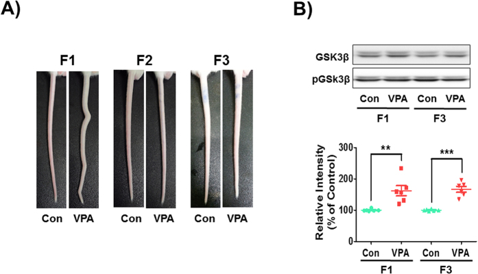
Figure 2. Neural tube defects by VPA exposure and its transgenerational effect.
(A) Malformations of tail structure occurred in F1 VPA-exposed mice, but not in the F2 and F3. (B) Protein levels of phosphorylated-GSK3β were examined using Western blot in F1 and F3 generation mice frontal cortex at embryonic day 14. All data are expressed as the mean ± S.E.M. (n = 12 mice randomly selected from 6 litters per group and per generation). **p < 0.01, ***p < 0.001 vs. control group as revealed by post-hoc Bonferroni’s comparisons.

